Digital laser microscope for deeper tissue
- Details
- Hits: 5147
With that of dr Catherine Philip developed digital Laser microscope can be in particular deeper layers of tissue examine how they z. B. found in patients in the thyroid gland. For her outstanding dissertation, Dr. Philipp with the Bertha Benz Prize 2020 honored.

Contents
So far in the Medical Technology many optical procedures at depths Tissue examinations reached its limits, because aberrations significantly impaired the image quality. At the professorship for measuring and sensor technology realized Dr. Philipp developed a new type of laser microscope with the support of the German Research Foundation (DFG).
Laser microscope penetrates deep tissue layers
She used one adaptive lens, which was developed at the University of Freiburg by the Professorship of Microactuators. For the first time, high-resolution images can be captured in deep tissue layers using minimalist adaptive optics. This allows for an easy-to-use and compact design.
The development also aims to democratize the computer-based laser beam microscope. “With this technology, a novel medical imaging procedurewhich, on the one hand, enables faster, simpler and more cost-effective diagnostics and, on the other hand, eliminates the need for complex chemical or mechanical preparation of the examined samples, ”says Dr. Philip.
Paradigm Shift in Biomedicine
The digital laser microscope opens up completely new diagnostic possibilities for the patient, especially when examining deeper tissue layers. It is used, for example, for examinations Changes in the thyroid gland. Dr. Phillip developed and built a laser microscope that represents a paradigm shift in biomedicine.
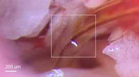
With her innovative laser microscope, Katrin Philipp recorded the fluorescence from the thyroid glands of zebrafish embryos. The animals did not have to be killed.
 Improved cochlear implant with 3D printing of microstructures
This species of fish represents a widespread model for the development and accumulation of harmful environmental toxins in the thyroid gland, including in humans.
In contrast to the previously laborious preparation of the samples after methodological gold standard with the subsequent cutting of the animals into thin slices, the measurement with the new technology takes place on one living organism under anesthesia. In principle, this allows the development over time to be followed on the same sample. In this way, it is possible to examine in more detail how environmental toxins affect the development of an organism.
Research work awarded the Bertha-Benz Prize
The new laser process is based on a theoretically and experimentally extremely demanding one research. With him an internationally visible progress was achieved for laser microscopy, which represents a special engineering achievement at the interface of biology, Microsystems Technology and Measurement Technology represents. The results of the research were published in the journal Scientific Reports released. The research work was done at the faculty Electrical Engineering and information technology of the TU Dresden submitted and with summa cum laude evaluated.
For your dissertation "Investigation of aberration correction and axial scanning in microscopy employing adaptive lenses" has the Daimler and Benz Foundation Dr. Katrin Philipp from the TU Dresden was awarded the Bertha Benz Prize 2020.
The 2020 Bertha Benz Prize winner
Dr. Katrin Philipp on her dissertation entitled Investigation of aberration correction and axial scanning in microscopy employing adaptive lenses: