 Robots can help doctors detect and treat tumors, for example by positioning a fine probe in the right place. So that Robotic imaging procedures such as magnetic resonance imaging, IPA engineers have developed a new drive technology.
Tumor therapy without risks and side effects? Still, this seems unthinkable. However, scientists in the laboratories are already working on solutions for the medicine of the future: "One of the major goals of research is to develop technologies for minimally invasive interventions that treat tumors so accurately and efficiently that no healthy tissue is destroyed." , explains Johannes Horsch from the project group Automation in Medicine and Biotechnology PAMB of the Fraunhofer IPA.
Position the probe using imaging
Together with his team, the engineer works on robots, with the help of which a surgeon can precisely position a fine probe, take a sample or thermally treat the tumor tissue. To bring such a probe exactly to the desired location requires skill and experience: If the doctor introduces the tiny probe with a needle, he must orient himself by means of images that indicate the position on the screen. "So far, X-ray-based methods are usually used for imaging. However, these have the disadvantage that they do not represent soft tissue, for example organs, very well. In addition, they cause an increased exposure to X-rays both in the doctor and in the patient, "explains Horsch.
Biggest problem: drive technology
"More potential for the future, therefore, magnetic resonance imaging, short MRI." Doctors who want to lead using MRI images a probe to a liver, lung or intestinal tumor quickly reach its limits: The tube in which the patient or the Patient is lying, leaves the surgeon hardly freedom of movement. To solve this problem, various research teams around the world are working on robots to help insert the needle. "The biggest problem is drive technology," reports Horsch. "The motors, we're talking about actuators, should not contain any ferromagnetic or electrically conductive materials as they can interfere with MRI imaging. Classic electric motors are therefore eliminated. "Even pneumatic cylinders that are difficult to control are not suitable.
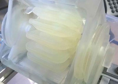
Plastic bellows as the heart of the drive
The solution of the IPA engineers: a hydraulic robot drive. At the heart of this actuator are plastic bellows made using 3D printing technology. These look like a little accordion connected to a thin, fluid-filled pipe. When the fluid is pressurized, the accordion expands or bends. This bend can be used to move a robotic arm that, for example, carries a needle probe.
By combining two hydraulic actuators, the robot arm can be precisely controlled in two spatial directions. Thanks to a force feedback mechanism, the surgeon who moves the robotic arm senses when the probe encounters resistance. "The real innovation is that the actuators do not contain any parts that interfere with the MRI recordings," concludes Horsch. The hydraulics can generate large forces in a small space. This solves the space problems within the MRI tube. Although you still need a motor that creates the pressure in the lines, but this can be well shielded in an adjoining room accommodate.
New drive technology meets expectations
Studies at the University Hospital Mannheim have now shown that the new drive technology meets expectations. "This laid the foundation for the development of a practical, robot-based positioning system for interventions on MRI," says Horsch. In a follow-up project, he and his team want to install the bending actuators in a robot that is also to be manufactured using 3D printing technology. The scientists and engineers want to test this in a preclinical study on replicas of human organs and tissues, as they are used for training of medical professionals.